Researchers usually study brain function by monitoring two types of electromagnetism: electric fields and light.
Currently, the most accurate way to monitor electrical activity in the brain is by inserting an electrode, which is very invasive and can cause tissue damage.
EEG is an easy method that uses simple technology to provide a good look into the brain, but measuring light emitted by luminescent proteins within the brain can be very challenging.
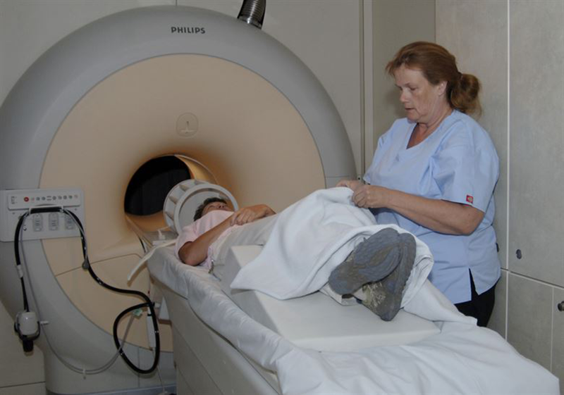
Now researchers at MIT have now adapted a new technique to detect either electrical activity or optical signals in the brain using a minimally invasive sensor for magnetic resonance imaging (MRI).
“MRI offers a way to sense things from the outside of the body in a minimally invasive fashion,” says Aviad Hai, an MIT postdoc and the lead author of the study. “It does not require a wired connection into the brain. We can implant the sensor and just leave it there.”
This type of sensor could give neuroscientists a spatially accurate way to identify electrical activity in the brain. It can also be used to measure light, and could be adapted to measure chemicals such as glucose, the researchers say, reports MIT.
The senior author of the paper is Alan Jasanoff, an MIT professor of biological engineering, brain and cognitive sciences, and nuclear science and engineering. Co-authors are: Virginia Spanoudaki and Benjamin Bartelle, both postdocs at MIT. The research was published at Nature Biomedical Engineering.
Prof. Jasanoff’s lab has previously developed MRI sensors that can detect calcium, serotonin and dopamine. In this new study, they wanted to expand their approach to detecting biophysical phenomena such as electricity and light.
The researchers built a tiny implantable antenna that is meant to work like the antenna built into the MRI machine.
The sensor is initially tuned to the same frequency as the radio waves emitted by the hydrogen atoms. When an electromagnetic signal from the tissue is detected by the sensor, its tuning changes and the sensor no longer matches the frequency of the hydrogen atoms. When this happens, a weaker image is produced when the sensor is scanned by an external MRI machine, reported MIT.
Read more Brain Advantage, The Wearable Device That May Improve Brain Performance
The researchers were able to show that the sensors can pick up electrical signals similar to those produced by the electrical impulses fired by single neurons, or the sum of electrical currents produced by a group of neurons.
The researchers are interested in using this type of sensor to detect neural signals in the brain, and they believe it could also be used to monitor electromagnetic phenomena in others parts of the body, such as muscle contractions or cardiac activity.


